

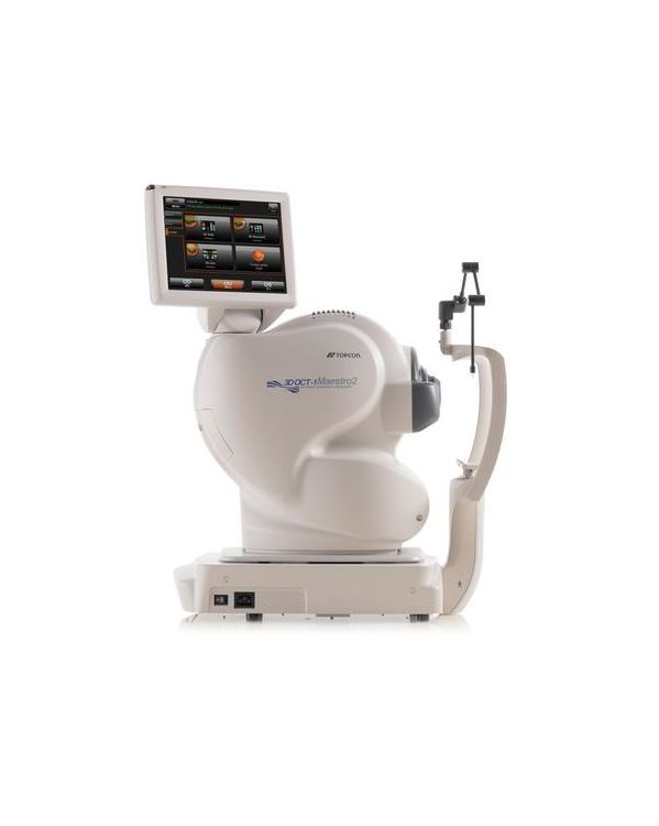
Topcon Maestro 2 - OCT and Fundus Camera *NEW*
Topcon Maestro 2 OCT and Fundus Camera
Topcon Maestro 2 OCT and Fundus Camera
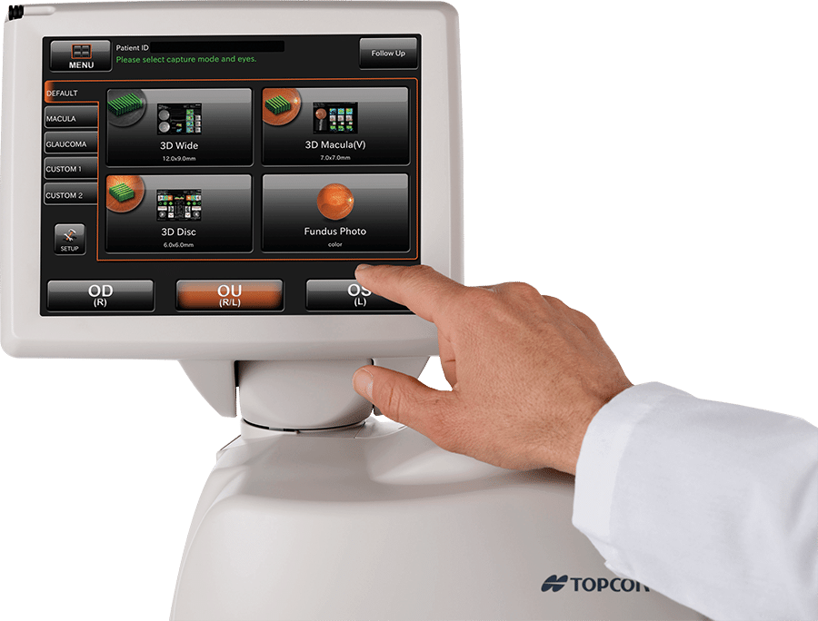
Maestro2 provides fast, multi-modality OCT/fundus imaging, and advanced data management. The complete clinical workstation for any busy practice, the Maestro2 automatically performs alignment, focus, optimizing and capture with a single touch. After capturing, the report can be immediately displayed by the simple click of an icon. In addition to automated capture, the Maestro2 offers manual/semi-manual options for difficult-to-image patients.
A fully automated OCT:
With one touch, you can simultaneously acquire a posterior OCT image and a true color fundus image.
This allows for PinPoint Registration and structural confirmation of the pathology. A small pupil function is also available, as well as fundus only capture.
Key Features
- Widefield OCT
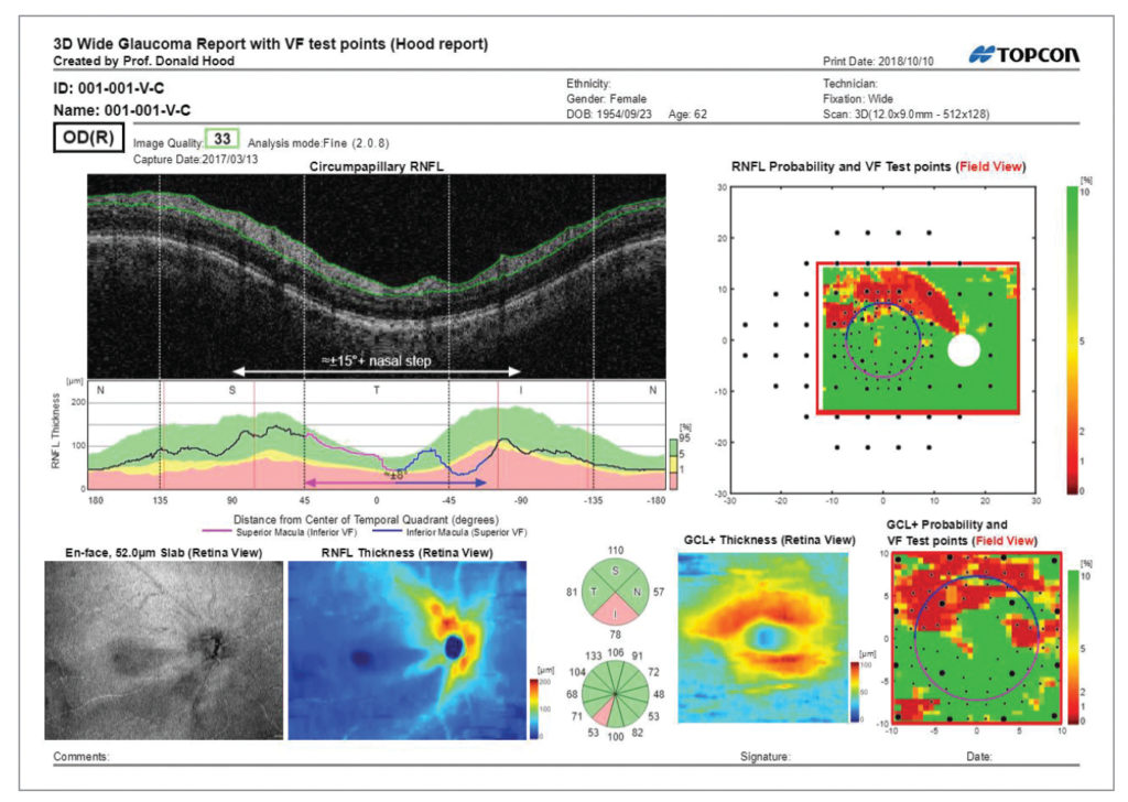
- The NEW Hood Report for Glaucoma
- Reference database for retina, RNFL, GCL+, and GCL++ thickness
- Panoramic fundus imaging
- Compact and space-saving design
- Combination OCT and true color fundus
- Automatic layer segmentation
- Anterior segment OCT
The Hood Report For Glaucoma
Developed in collaboration with Professor Donald C. Hood, the Hood Report for Glaucoma aids understanding of the relationship between retinal damage and visual field loss.
HIGH RESOLUTION OCT AND COLOR FUNDUS PHOTOGRAPHY
A high-resolution B-scan and smooth 3D graphics facilitate the observation of pathology and each layer of the retina. High-quality color fundus photography gives fundamental and additional information. The OCT and color fundus are an inseparable combination for daily diagnosis.
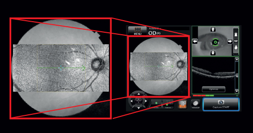
PERIPHERAL FUNDUS PHOTOGRAPHY
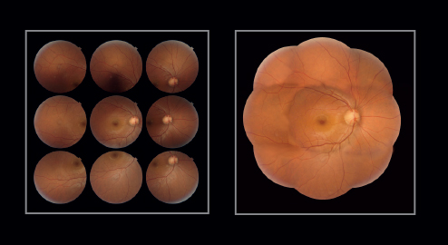
The Maestro2 allows the operator to automatically select 9 standard fields or manually manipulate the patient’s fixation to create a mosaic image with the AutoMosaic software.
LIVE VIEW FUNDUS™
OCT-LFV is a live projection image of the retina. The clear live fundus image makes the disc, retinal vessel and scanning position easy to see, when required.
CATARACT MODE
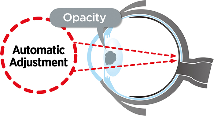
Cataract mode automatically moves the scanning position on the upper/lower (or L/R) area to adjust for opacity in the eye due to cataract.